

 View Portfolio |
 Inside Time |
 Shows |
 About/Contact the Artist |
The Sense of Taste.Scientists are on the verge of great breakthroughs in how taste works. Some information about the structures of the proteins which receive the different tastes is known, but the full structures have not yet been solved. Older ideas about locatoins of the different taste sensations on the toungue are changeing. It is also now believed that some of us may taste differently than others, due to different variantions in each person's taste receptor genes.
See "Making Sense of Taste" by David V. Smith and Robert F. Margolskee, Scientific American, March 2001. Also, "In the realm of your senses", New Scientist, January 31 - February 6, 2004.
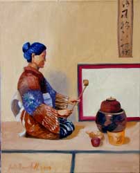
"Study for Taste Bud Kimono for a Japanese Tea Ceremony", 16" x 20"(left); Japanese Tea Ceremony with Taste Bud Kimono", 16" x 20" (right), 2003
Oil and mixed media (left) and Oil on Canvas (right). $1,600 for the pair.
These are small, prototype paintings. There will be larger, 30" x 40" versions of this work, as well as a series of real Kimonos. As the Japanese Tea Ceremony emphasizes the use of all the senses, there will be a Kimono designed for each taste, using imagery of the types of cells and receptors involved in that particular taste.
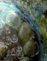
Taste bud images courtesy of Bernd Lindemann, University of Saarlandes, from the paper "Multi-photon microscopy of cell types in the viable taste disk of the frog", Jack H.-Y. Li · Bernd Lindemann, Cell Tissue Res (2003) 313:11–27.
)
"A Taste of India Tablecloth" (left) 30" x 40"; "Ayurvedic Still Life"(right) 33" x 44", 2003
Prints available
According to the very old Ayurvedic tradition in India, it is important to properly balance the different kinds of tastes in a meal. There are traditionally six different kinds of taste in this practice; sweet, sour, salty, pungent, bitter and astringent. Each taste is important for different organs and different aspects of health. If one is in balance, they would optimally have some of each taste in their meals. This still life is arranged according to these tastes. Sweet is on the left, Jaggery and sugar crystals, then a lemon and green mango for sour, a box of Asafetida, a chili pepper, ginger and onions, and some cardamon for pungent, a bottle of salt, bitter melon, and astringent flavors at the end including green bananas, yellow split peas, tumeric and raw vegetables.
Scientific Credits:
At the corners of the rug are models of the sweet receptor, based on mGluR1, from the labs of Robert Margolski and Marianna Max, created by Marianna Max. A very sweet molecule, brazzein, which binds to the sweet receptor, surrounds the sweet receptor at the corners. The blue and green protein structures at the edges are the open and closed form of mGluR1, the receptor for glutamate, or the umami receptor. These I created using the Crystal Structure Of Metabotropic Glutamate Receptor Subtype 1 Complexed With Glutamate (1EWK) N.Kunishima, Y.Shimada, H.Jingami & K.Morikawa, and without (1EWT). Glutamate runs along the borders of these proteins. These I made with UCSF Chimera software. Underneath the glutamate are images of images of cells stained for the bitter receptor by Dr. Nick Ryba. In the center of the image is a microscope image of a taste bud from a fish by Anne Hansen. Models of bitter receptors are in golden at the corners of the central portion of the rug. They were modeled after the rhodopsin receptor by Marianna Max. The clusters of four white circular shapes around the bitter receptor are representations of how the sour receptor proteins might arrange themselves, in groups of four. The larger, eight circle flower like shapes around the taste bud are possible arrangements of the salty receptor proteins. In this case, the central port formed by the eight proteins would be where the sodium ions were allowed to enter. This information was provided by Candice Askwith, and the original paper on the ENaC Epithelium Sodium Channel came from J Biol Chem, Vol. 274, Issue 38, 27281-27286, September 17, 1999, Eskandari, et. al
The view through the window is an adaptation of an electron microscope image of a developing human taste bud taken by Martin Witt of the University of Technology Dresden.Other credits:
"Textbook of Ayurveda Fundamental Principles, Volume One" by Vasant Lad, M.A.Sc., The Ayurvedic Press, 2002, ISBN 1-883725-07-0.
"Indian Food, A Historical Companion", K.T. Achaya, Oxford University Press, 1994.
"Textile Arts of India", Kokyo Hatanaka Collection, 1993, ISBN 0-8118-1084-4
)
)
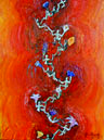
"Kuba Tablecloth", 30" x 40";
Series of two IN PROGRESS Shoowa of Kuba, Africa.
The Kuba people in central Africa are known for their beautiful and sophisticated textiles. They are either made on a raphia panel (a type of fiber grown in the region), tied and stitch-resist, or on bark. Nothing is pre-designed or drawn ahead of time. The design evolves as it is made.
This design incorporates taste bud images, and was composed in a way which reminded me of the Kuba designs.
The the still life panel with the textile and the food of this region are in progress as well and no sketch is yet available.
See "African Textiles" by John Gillow, Thames and Hudson Limited, ISBN 0-8228-4266-9. Taste bud images courtesy of Anne Hansen.
)