
...Julie Newdoll merges life science and culture, myths and molecules in her
paintings, music, journal covers and science games.
Shakespeare:
A Mirror up to Science
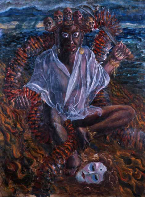
Manjushri as topoisomerase, Untier of DNA Knots, oil on canvas,
3' x 4', 1997. Private collection.
Topisomerase is a protein that is found inside the cells in our bodies, inside the nucleus. When a cell is in between divisions, the DNA of the cell is relaxed, looking much like a bowl of spaghetti. When it is time for the cell to divide, the DNA tightens up into its respective chromosome. Sometimes, a knot has formed in a strand of DNA, preventing it from moving where it needs to go. This protein detects such knots, cuts the DNA at the knot, unties the knot, and attaches the DNA ends back together. Manjushri is, in part, a God of knowledge. His sword cuts through untruths, and heals on the way out.
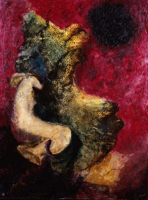
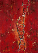
Woman Bound to DNA I, 30" x 40", 1992. $8,000, Available for purchase. Prints available $450.
The shape of DNA contains a major groove and a minor groove. You can see the major groove in the center of the image, with a saddle shape. The figure of a woman is bound to the structure. This type of surface representation of DNA is called a "solvent accessible surface", as it indicates how close water molecules could get to the DNA. A computer generated image of DNA was first applied to the canvas using photo-emulsion, and the painting was done on top of that.
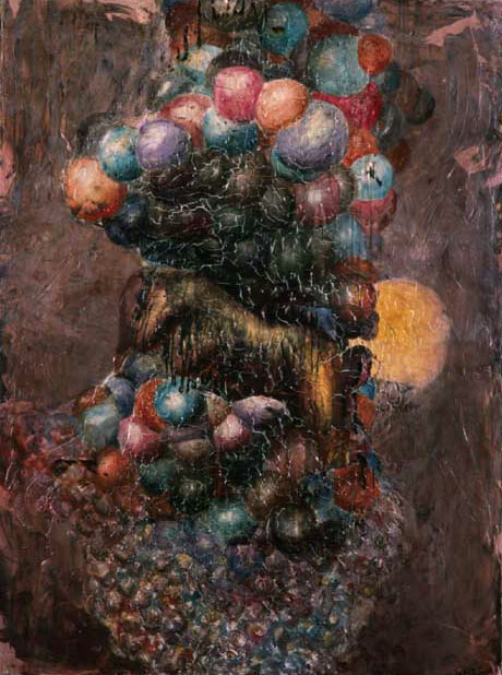
Woman Bound to DNA II, 30" x 40", 1992. $8,000, Available for purchase.
The shape of DNA contains a major groove and a minor groove. You can see the major groove in the center of the image, with an indent near the yellow sphere. The figure of a woman is bound in the major groove. The atoms of the DNA are represented by spheres. A computer generated image of DNA was first applied to the canvas using photo-emulsion, and the painting was done on top of that.
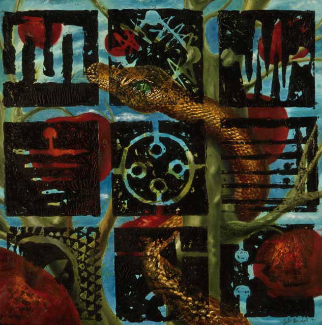
Knowledge, oil on canvas, 38" x 38", 1991, private collection.
Special thanks to my father for consultation about the electrical symbols.
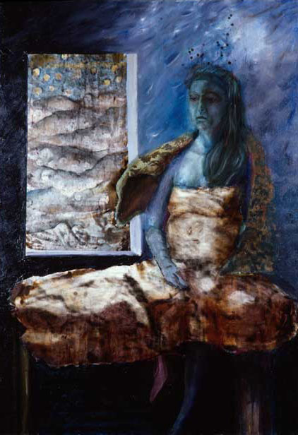
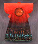
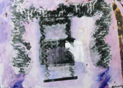
Robin in a Sperm Surface Dress, oil on canvas and mixed media, 4' x 5', 1994. Available for purchase. $750 unframed. Prints available $450.
The patterns in the window and on the dress are from electron microscope images of the surface of a sperm. Electron Microscope Images supplied by Dr. Dan Friend, Harvard Medical School, Pathology Department.

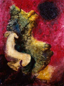
Spermbrella for Black Days, oil on canvas, 4' x 5', 1994. $800. Prints available $450.
The sickle-like shape is taken from an electron microscope image of a guinea pig sperm. Microscope image supplied by Dr. Dan Friend, Harvard Medical School, Pathology Department
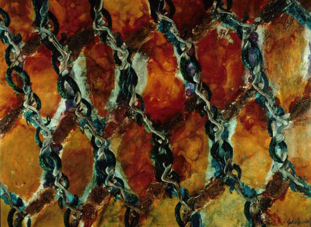
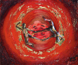
Lilith in a Colicin Crystal, oil and mixed media on canvas, 3' x 4', 1996, Collection of Professor Robert M. Stroud. Prints available $450.
A protein crystal structure composed of Lilith figures. Her arms, hair, and serpent tail form a relationship like that of the Colicin IA molecule when it is crystallized. Colicin is a long protein harbored by bacteria. A single molecule of it can be directed at a neighboring bacteria and kill it with a kind of punture wound. Lilith has similar potency.
Consultation for painting science with Robert M. Stroud. Paper: "Crystal structure of colicin Ia.", Wiener M; Freymann D; Ghosh P; Stroud RM , Nature 1997 Jan 30; 385(6615):461-4
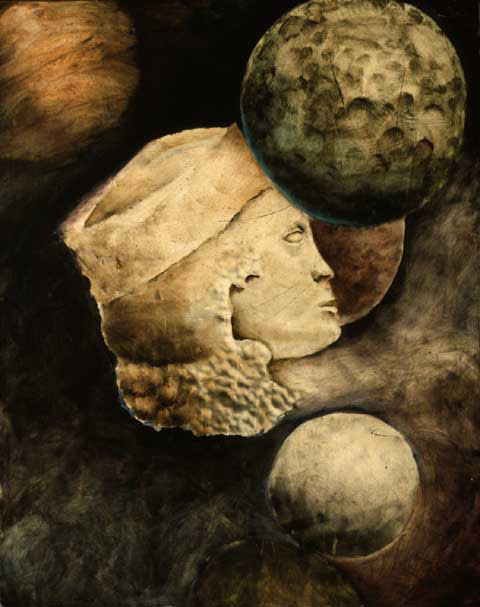
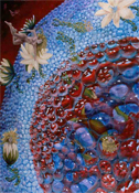
Microantiquity, oil and mixed media, 30" x 38", 1993, private collection.
Beginning with an electron microscope image of pits on the surface of a cell, this painting evolved. Microscope image supplied by Dr. Dan Friend.
Programmed
Cell Death:
Apoptosis

Copyright 2012 Julie Newdoll
